
Bone fracture systems
_____
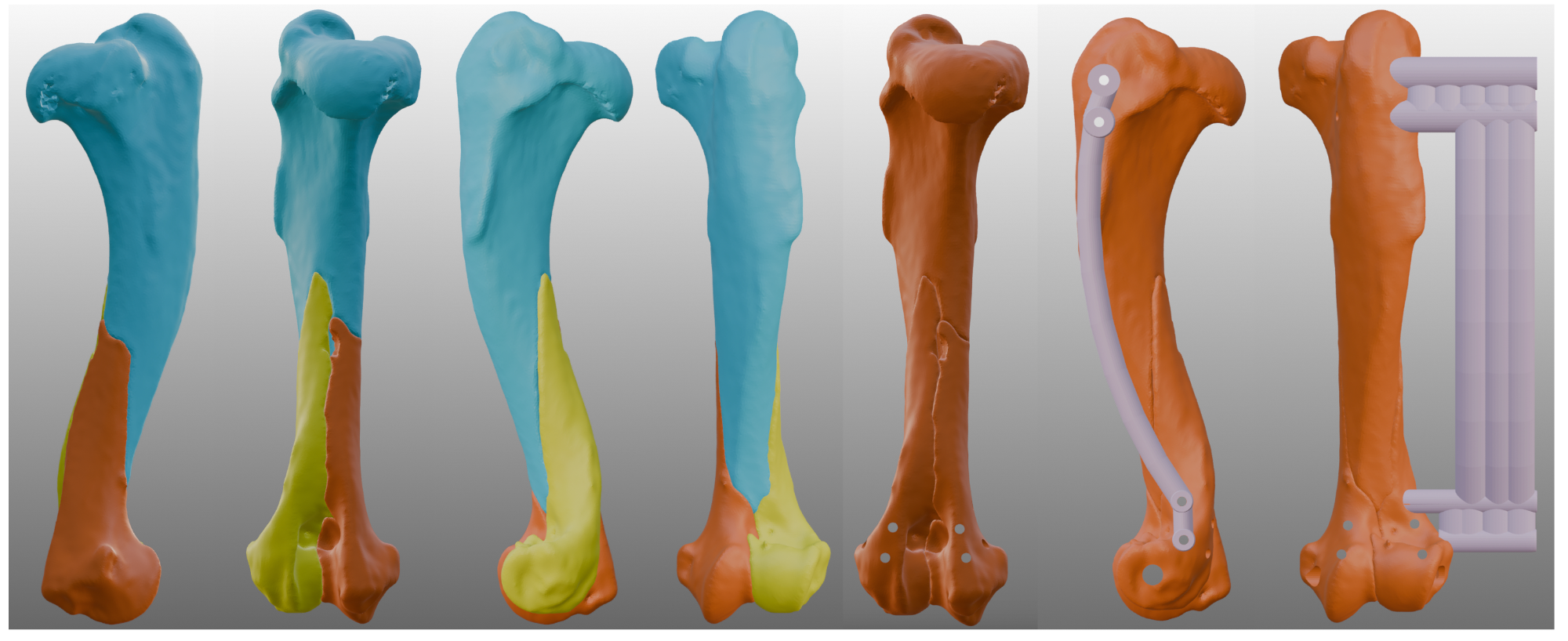
Complex fractures present some of the most demanding scenarios in orthopaedic surgery. Multiple fragments, loss of normal landmarks, and the difficulty of visualising the intended reduction can make these repairs technically challenging and time-consuming. A patient-specific approach — combining a printed bone model with custom guides and jigs where appropriate — can simplify these procedures and bring greater predictability to both planning and execution.
1. Simplifying complex repairs, including minimally invasive surgery
Where a fracture involves multiple fragments or distorted anatomy, planning the reduction in advance on a three-dimensional model allows the reconstruction to be worked out before surgery rather than during it. The intended fragment positions and fixation strategy are established beforehand, which can take much of the uncertainty out of a difficult repair and provide a clear, predetermined plan to follow in theatre. Custom reduction guides and jigs can then be designed to hold the fragments in their planned positions during implant fixation, maintaining the reduction at the critical moment when it is most easily lost.
This planning is particularly valuable in minimally invasive approaches, where direct visualisation of the fracture is deliberately limited to preserve the soft-tissue envelope and biology of healing. With the reduction and implant position defined in advance — and the implant pre-contoured to fit — the repair can be carried out through smaller approaches with greater confidence, reducing reliance on extensive exposure and repeated intra-operative imaging. Here, reduction jigs are especially useful, securing the fragments in the planned alignment with minimal additional exposure while fixation is applied.
2. Plate pre-contouring
Reducing the fracture virtually produces a model of the restored anatomy against which plates and other implants can be shaped before surgery. Pre-contouring in this way means the implant is ready for immediate use, improves the accuracy of fit to the reconstructed bone, reduces intra-operative handling time, and limits the repeated bending that can weaken an implant. Where available and appropriate, the contralateral limb can also serve as a surgical reference and be printed as a mirror-image model, providing an intact anatomical template against which the implant can be pre-contoured.
3. Anatomy referencing
A sterilisable model of the intended reconstruction provides a tangible intra-operative reference to the target anatomy. This is especially helpful where landmarks are obscured or distorted by the fracture, supporting orientation and decision-making throughout the procedure.